
Sonography, also known as ultrasound imaging, is a diagnostic tool used in gynaecology to produce images of the reproductive system. It uses high-frequency sound waves to create images of the uterus, ovaries, and other pelvic organs, allowing physicians to visualize and evaluate the structure and function of these organs.
Sonography is a non-invasive and painless procedure that is often used to diagnose and monitor a variety of gynaecological conditions, such as fibroids, ovarian cysts, endometriosis, and uterine abnormalities. It can also be used to monitor pregnancies and assess fetal development.
During a gynaecological sonography, the patient lies on an examination table, and a small wand, called a transducer, is placed on the abdomen or inserted into the vagina. The transducer emits high-frequency sound waves that penetrate the body and bounce back to create images on a computer screen.
Sonography in gynaecology is a safe and effective diagnostic tool that can provide valuable information to our best gynaecologist in Surat about the health and function of the reproductive system. It is often used in conjunction with other diagnostic tests, such as blood tests and biopsies, to provide a comprehensive evaluation of gynaecological conditions.
At Ookid Hospital & IVF Centre, the best sonography centre in Surat, we provide a range of high-quality ultrasound imaging services to help diagnose and monitor a variety of medical conditions. Our team of experienced and skilled sonographers uses state-of-the-art equipment to produce detailed images of internal organs and tissues.
Our sonography services include a wide range of diagnostic imaging tests, including abdominal, pelvic, obstetric, gynecologic, breast, and musculoskeletal sonography. We use the latest technology and techniques to provide accurate and detailed images that can help physicians make informed diagnoses and treatment decisions.
Our sonographers are trained and experienced in using the latest ultrasound technology to produce high-quality images quickly and efficiently. They work closely with gynaecologist to ensure that the imaging studies are tailored to meet the specific needs of each patient.
At our sonography centre, we understand that undergoing medical imaging tests can be stressful and intimidating. That’s why we prioritize patient comfort and care throughout the entire imaging process. Our team is dedicated to ensuring that patients feel comfortable and informed throughout the entire process, from check-in to receiving their results.
In addition to our advanced imaging technology and compassionate care, we also offer affordable pricing and convenient scheduling options. We accept most insurance plans and offer competitive pricing for self-pay patients. We also offer flexible scheduling options to accommodate our patients’ busy schedules.
If you’re looking for the best sonography centre in Surat, look no further than Oorkid Hospital & IVF Centre. Our team of experienced and skilled sonographers is dedicated to providing the highest quality imaging services to our patients. We use the latest technology and techniques to provide accurate and detailed imaging studies that can help physicians make informed diagnoses and treatment decisions.
Whether you’re in need of routine imaging studies or specialized diagnostic tests, our sonography centre is here to provide you with the care and support you need. Contact us today to schedule an appointment and experience the difference that our advanced technology and compassionate care can make.
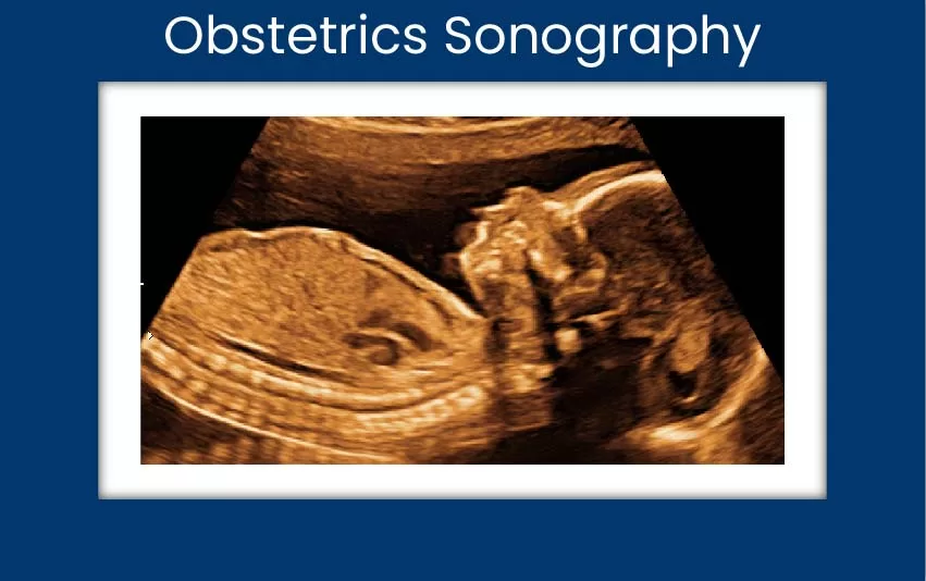
A center for obstetric sonography, also known as a prenatal ultrasound center, is a specialized facility that provides ultrasound services to expectant mothers during pregnancy. These services include routine prenatal scans and more detailed evaluations to assess the growth, development, and health of the fetus.
At Orkid hospital, our center for obstetric sonography is staffed by highly trained and experienced sonographers and obstetricians who use state-of-the-art ultrasound equipment to produce high-resolution images of the fetus. Our center offers a wide range of ultrasound services, including:
Routine obstetric scans: These scans are performed at regular intervals throughout pregnancy to monitor the growth and development of the fetus, assess the position of the placenta, and check for any abnormalities or complications.
Detailed obstetric scans: These scans are performed at specific times during pregnancy, such as in the first trimester, to assess the health of the fetus in more detail and check for any chromosomal abnormalities or structural defects.
3D and 4D ultrasound: These types of ultrasound create three-dimensional and four-dimensional images of the fetus, respectively, which can provide more detailed images of the fetus’s face, hands, and feet.
Obstetric sonography is a safe and non-invasive procedure that is widely used to monitor the health and development of the fetus during pregnancy. At Orkid hospital, our center for obstetric sonography is dedicated to providing expectant mothers with the highest level of care and support throughout their pregnancy.
If you are pregnant and have been referred for obstetric sonography, you can expect the highest quality care at Orkid hospital. For more information, mail us at info@orkidmedilife.com

Infertility sonography, also known as reproductive ultrasound, is a diagnostic tool used to evaluate reproductive organs and diagnose causes of infertility. The procedure uses high-frequency sound waves to create images of the uterus, ovaries, and fallopian tubes, which are then used to assess the condition of the reproductive organs and identify any abnormalities that may be causing infertility.
At Orkid hospital, our infertility sonography services are performed by trained and experienced sonographers who use state-of-the-art ultrasound equipment to produce high-resolution images. The procedure is typically performed on an outpatient basis and does not require any anesthesia.
There are several types of infertility sonography, including:
Infertility sonography can help diagnose conditions such as polycystic ovary syndrome (PCOS), endometriosis, fibroids, and pelvic inflammatory disease (PID), as well as identify problems with ovulation or sperm quality.
If you and your partner are experiencing difficulty conceiving, then Orkid’s sonography with the latest technology-equipped equipment can help guide your treatment options and increase your chances of having a baby.
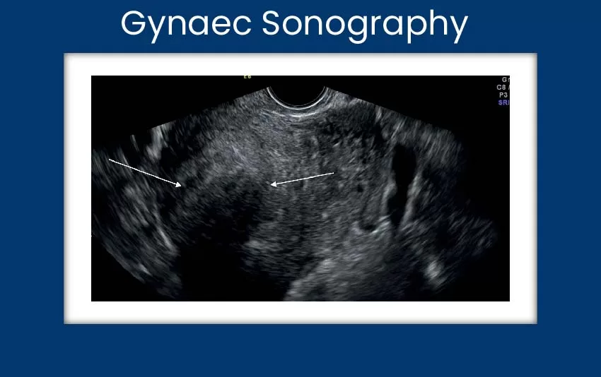
Gynecological sonography, also known as gynecological ultrasound, is a diagnostic procedure that uses high-frequency sound waves to create images of the female reproductive organs, including the uterus, ovaries, and fallopian tubes. The procedure is used to assess the condition of the reproductive organs and identify any abnormalities that may be causing gynecological problems.
At Orkid hospital, our gynecological sonography services are performed by trained and experienced sonographers who use state-of-the-art ultrasound equipment to produce high-resolution images. The procedure is typically performed on an outpatient basis and does not require any anesthesia.
There are several types of gynecological sonography, we offer at our hospital, some of which are listed below:
At Orkid hospital gynaec sonography is generally safe and well-tolerated, and the results can help guide your treatment options and ensure the best possible outcome.
Get insider tips straight to your inbox, relate to the challenges other services teams face everyday.
© 2025 Orkid Hospital & IVF Centre. All Rights Reserved.
Powered By